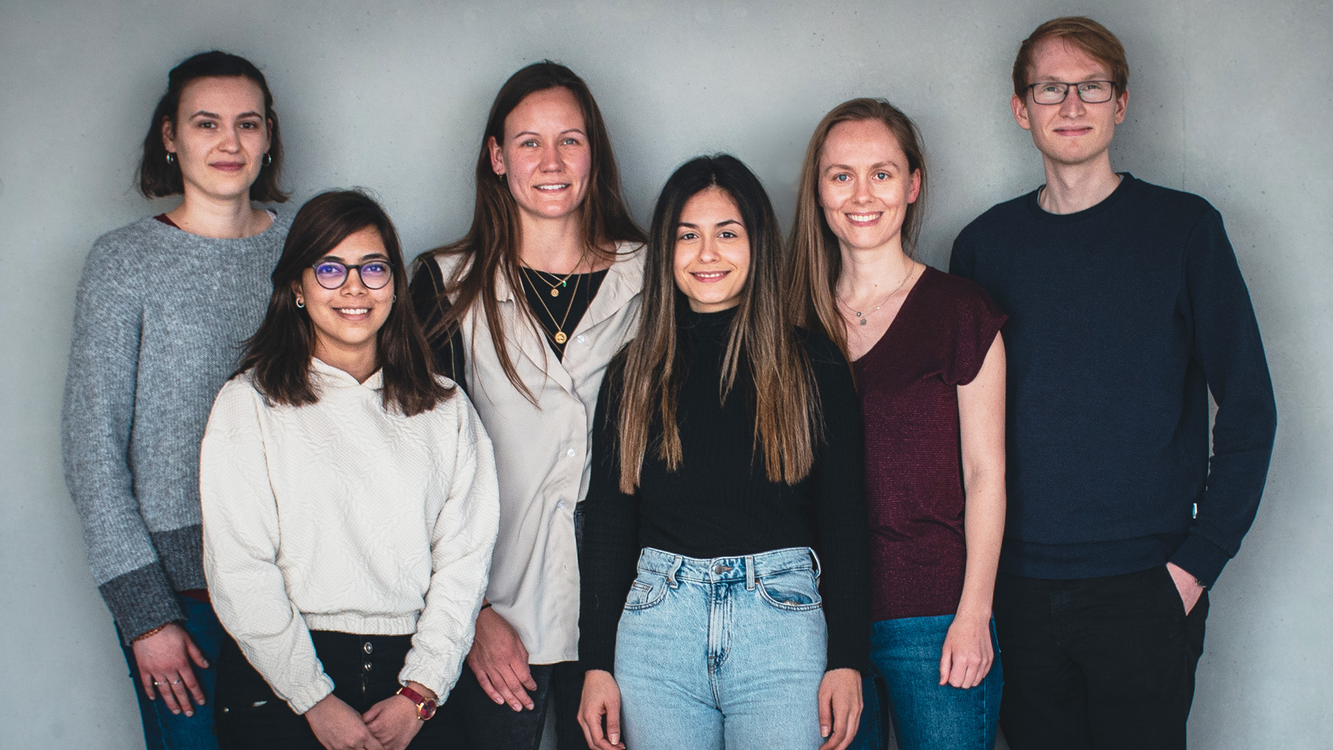
Antigen-specific T and B cells are the two key players of the adaptive immune response. The interaction between these two cell types has to be balanced to guarantee a successful protective immune response but to prevent the development of autoimmune disease. Our lab investigates the molecular and cellular regulation of T/B cell interaction in secondary lymphoid organs and chronically inflamed tissues.
Group leader:
,
Group members:
,
,
,
,
,
,
,
The interaction of antigen-specific T and B cells is key for an effective adaptive immune response. T follicular helper (Tfh) cells are the T cell subset that provide B cell assistance during the germinal center reaction. Tfh cells are the prerequisite for the generation of high-affinity memory B cells and long-lived plasma cells. Therefore, manipulation of the Tfh response is of particular clinical interest for aiding in the generation of protective antibodies during vaccination or for eliminating harmful antibodies in autoimmune diseases and allergies [1].
Our group’s main focus is on the generation of Tfh cells, their maintenance, and autoimmune diseases resulting from a dysregulated germinal center response. We previously identified several key regulators of Tfh cells including the T cell costimulator ICOS [2] and the transcription factors Klf2 [3] and Bach2 [4]. We are currently mapping the very early events of Tfh cell differentiation in vivo to develop in cooperation with Kevin Thurley (University of Bonn) an in silico model that presents how different cytokines influence the differentiation of T helper subsets.
T cell/B cell cooperation normally takes place in secondary lymphoid organs. However, we recently identified a novel population of Tfh-like cells, now also known as peripheral T helper cells (Tph), which drives B cell differentiation in inflamed non-lymphoid tissues [5]. Using a mouse lung inflammation model, we want to elucidate further how T/B interaction can take place outside the ordered structure of the germinal center normally present in secondary lymphatic organs.
These Tfh-like cells can also be found in the lung of human sarcoidosis patients [6]. They promote the local differentiation of plasma blasts directly in the inflamed lung tissue. These findings highlight a so far underappreciated role of B cells in the pathogenesis of sarcoidosis. We are currently analyzing T cell / B cell cooperation in non-lymphoid tissues from different chronic inflammatory conditions like pemphigoid disease.
To analyze the interaction of T and B cells in vivo , our group developed an adoptive transfer mouse model which allows to track antigen-specific T and B cells using multiparametric flow cytometry, confocal laser scanning microscopy, and single cell RNA sequencing [7]. Moreover, T and B cells can be manipulated using a retroviral overexpression system or CRISPR/Cas9-mediated knock-out to study the function of individual genes [4].
References
[1] Ritzau-Jost J, Hutloff A. T Cell/B Cell Interactions in the Establishment of Protective Immunity. Vaccines (2021) 9:1074.
[2] Hutloff A, Dittrich AM, Beier KC, Eljaschewitsch B, Kraft R, Anagnostopoulos I, Kroczek RA. ICOS is an inducible T-cell co-stimulator structurally and functionally related to CD28. Nature (1999) 397:263-266.
[3] Weber JP, Fuhrmann F, Feist RK, Lahmann A, Al Baz MS, Gentz LJ, Vu Van D, Mages HW, Haftmann C, Riedel R, Grün JR, Schuh W, Kroczek RA, Radbruch A, Mashreghi MF, Hutloff A. ICOS maintains the T follicular helper cell phenotype by downregulating Krüppel-like factor 2. J Exp Med (2015) 212: 217-233.
[4] Lahmann A, Kuhrau J, Fuhrmann F, Heinrich F, Bauer L, Durek P, Mashreghi MF, Hutloff A. Bach2 Controls T Follicular Helper Cells by Direct Repression of Bcl-6. J Immunol (2019) 202:2229-2239
[5] Hutloff A. T follicular helper-like cells in inflamed non-lymphoid tissues. Front Immunol (2018) 9:1707.
[6] Bauer L, Müller LJ, Volkers SM, Heinrich F, Mashreghi MF, Ruppert C, Sander LE, Hutloff A. Follicular helper-like T cells in the lung highlight a novel role of B cells in sarcoidosis. Am J Respir Crit Care Med (2021) 204:1403-1417.
[7] Bauer L, Hutloff A. Analysis of T-Cells in Inflamed Nonlymphoid Tissues. Methods Mol Biol (2021) 2285: 91-98.
University Hospital Schleswig-Holstein
Institute of Immunology
Arnold-Heller-Straße 3
Haus U30
24105 Kiel